Home » Uncategories » Compact Bone Diagram : Compact Bone Definition And Function Biology Dictionary / The cells of compact bone, which is also called cortical bone, appear to be tightly packed into a solid mass.
Friday, 18 June 2021
Compact Bone Diagram : Compact Bone Definition And Function Biology Dictionary / The cells of compact bone, which is also called cortical bone, appear to be tightly packed into a solid mass.
Compact Bone Diagram : Compact Bone Definition And Function Biology Dictionary / The cells of compact bone, which is also called cortical bone, appear to be tightly packed into a solid mass.. The compact bones form the hard exterior of the bones, whereas the spongy bones have several pores that are filled with nerves and blood vessels. To the naked eye, the compact bone is a solid layer present as the external layer of all bones. The cells of compact bone, which is also called cortical bone, appear to be tightly packed into a solid mass. Compact bone, also called cortical bone, is the hard, stiff, smooth, thin, white bone tissue that surrounds all bones in the human body. Compact bone is made of a matrix of hard mineral salts reinforced with tough collagen fibers.
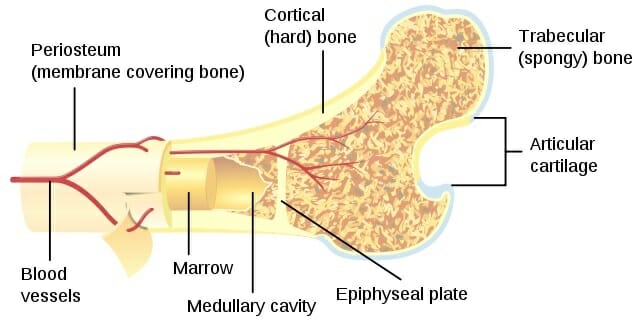
Some, mostly older, compact bone is remodelled to form these haversian systems (or osteons). Anatomy of a long bone proximal epiphysis diaphysis distal epiphysis compact bone spongy bone medullary cavity. (on textbook page diagrams note only highlighted labels) compact bone. Deep to the compact bone layer is a region of spongy bone where the bone tissue grows in thin columns called. Because of its strength, the compact bone makes it possible for the bone to support weight.
Art Labeling Activity Structure Of Compact Bone Diagram Quizlet from o.quizlet.com Add to favorites 0 favs. Diagram of a typical long bone showing both cortical (compact) and cancellous (spongy) bone. Online quiz to learn compact bone diagram; (b) in this micrograph of the osteon, you can clearly see the concentric lamellae and central canals. The compact bones form the hard exterior of the bones, whereas the spongy bones have several pores that are filled with nerves and blood vessels. The light spot is a canal that carries a blood vessel and a nerve fiber. They allow blood vessels and nerves to travel through them to supply the osteocytes. It is also called osseous tissue or cortical bone and it provides structure and support for an organism as part of its skeleton, in addition to being a location for the storage of minerals like calcium.about 80% of the weight of the human skeleton comes from.
Important for compression, especially at joints.
There are small canals that run through the bone, which allow blood vessels to penetrate it. Concentric lamellae interstitial lamellae central canal lacuna osteocyte canaliculus. Many tiny cells called osteocytes live in small spaces in the matrix and help to maintain the strength and integrity of the compact bone. Other sets by this creator. Osteon model lacunae canaliculi osteocyte. There are pores and spaces even in compact bone. (on textbook page diagrams note only highlighted labels) compact bone. You need to get 100% to score the 15 points available. External layer of all bone. Compact bone accounts for 80% of the bones in the human body. To the naked eye, the compact bone is a solid layer present as the external layer of all bones. Online quiz to learn compact bone diagram; Compact bone, also called cortical bone, dense bone in which the bony matrix is solidly filled with organic ground substance and inorganic salts, leaving only tiny spaces (lacunae) that contain the osteocytes, or bone cells.compact bone makes up 80 percent of the human skeleton;
Each osteon looks like a ring with a light spot in the center. 100x on this image you can see several of the structural units of bone tissue (osteons or haversian systems). Provides protection and support while resisting stress from weight and movement. Compact bone, as opposed to spongy bone, is made of cylindrical units, called osteons, that are tightly formed together. Compact bone is formed from a number of osteons, which are circular units of bone material and blood vessels.
Compact Bone Definition And Function Biology Dictionary from biologydictionary.net Diagram of a typical long bone showing both cortical (compact) and cancellous (spongy) bone. Compact bone is solid and has greater structural strength. In long bones, as you move from the outer cortical compact bone to the inner medullary cavity, the bone transitions to spongy bone. Elements with symbols from latin. You need to get 100% to score the 15 points available. Deep to the compact bone layer is a region of spongy bone where the bone tissue grows in thin columns called. They allow blood vessels and nerves to travel through them to supply the osteocytes. Some, mostly older, compact bone is remodelled to form these haversian systems (or osteons).
Deep to the compact bone layer is a region of spongy bone where the bone tissue grows in thin columns called.
To the naked eye, the compact bone is a solid layer present as the external layer of all bones. A diagram of the anatomy of a bone, showing the compact bone. Compact and spongy.the names imply that the two types differ in density, or how tightly the tissue is packed together. Bone connective tissue there are two types of bone tissue: The diagram above shows a longitudinal view of an osteon. It is dense (because of calcified matrix) with tiny spaces known as lucanas. Learn vocabulary, terms, and more with flashcards, games, and other study tools. (b) in this micrograph of the osteon, you can clearly see the concentric lamellae and central canals. The light spot is a canal that carries a blood vessel and a nerve fiber. Diagram of a typical long bone showing both cortical (compact) and cancellous (spongy) bone. (b) in this micrograph of the osteon, you can clearly see the concentric lamellae and central canals. Provides protection and support while resisting stress from weight and movement. The compact bones form the hard exterior of the bones, whereas the spongy bones have several pores that are filled with nerves and blood vessels.
The remainder of the bone is formed by cancellous or spongy bone. Found in short bones, flat bones, irregular bones, and end of long bones. Online quiz to learn compact bone diagram; Add to favorites 0 favs. The diagram above shows a longitudinal view of an osteon.
Lamellar Bone An Overview Sciencedirect Topics from ars.els-cdn.com You need to get 100% to score the 15 points available. A diagram of the anatomy of a bone, showing the compact bone. Because of its strength, the compact bone makes it possible for the bone to support weight. The diagram above shows a longitudinal view of an osteon. The cells of compact bone, which is also called cortical bone, appear to be tightly packed into a solid mass. Under periosteum of all bones is the bulk of the diaphysis of long bones. Terms in this set (8) spongy bone (contains red marrow) compact bone (has osteons) osteon. Related posts of compact bone diagram labeled anatomical diagram of internal organs.
The diagram above shows a longitudinal view of an osteon.
Serves as protection of bone marrow. Compact and spongy.the names imply that the two types differ in density, or how tightly the tissue is packed together. Each osteon looks like a ring with a light spot in the center. Compact bone is the strongest form of bone tissue containing few spaces. You need to get 100% to score the 15 points available. There are pores and spaces even in compact bone. This is most common and has the appearance of the matrix forming concentric rings around cavities for blood vessels. Important for compression, especially at joints. Provides protection and support while resisting stress from weight and movement. The compact bones form the hard exterior of the bones, whereas the spongy bones have several pores that are filled with nerves and blood vessels. Compact bone, also called cortical bone, is the hard, stiff, smooth, thin, white bone tissue that surrounds all bones in the human body. They allow blood vessels and nerves to travel through them to supply the osteocytes. Haversian canals (sometimes canals of havers) are a series of microscopic tubes in the outermost region of bone called cortical bone.
0 Response to "Compact Bone Diagram : Compact Bone Definition And Function Biology Dictionary / The cells of compact bone, which is also called cortical bone, appear to be tightly packed into a solid mass."


0 Response to "Compact Bone Diagram : Compact Bone Definition And Function Biology Dictionary / The cells of compact bone, which is also called cortical bone, appear to be tightly packed into a solid mass."
Post a Comment